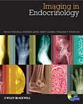
Imaging in Endocrinology will provide endocrinologists and
radiologists of all levels with an outstanding diagnostic imaging
atlas to aid them in the diagnosis and management of all the
major endocrine diseases they are likely to encounter.
In full colour throughout, the 300 high-quality images
consist of CT scans, MRI, NMR and histopathology slides,
and are arranged by each specific endocrine condition, resulting in
a visually outstanding and easily accessible tool that guides
the user through exactly what to look out for and provides a
practical and extremely useful aid in helping them formulate a
diagnosis.
Every major endocrine condition is covered in a specific
section, including diseases of the thyroid, pituitary,
reproductive and adrenal glands, the pancreas, bone metabolism
problems, and the various forms of endocrine
cancers. Each disease covered will offer a comparison of
the normal findings so as to further assist
in diagnosis. An accompanying website contains an
online slide-atlas of all the figures in the book, to
allow users to download all figures for use in
presentations.
Led by Paolo Pozzilli, an internationally-recognised expert in
this field, the authors have assembled a wonderful collection of
images that will be greatly valued by endocrinologists and
radiologists alike, ensuring this is the perfect tool to
consult when assessing patients with endocrine disease.
Autorentext
Paolo Pozzilli MD
Department of Endocrinology & Diabetes, University Campus Bio-Medico di Roma, Rome, Italy; Centre of
Diabetes, The Blizard Institute, St. Bartholomew's and the London School of Medicine, Queen Mary
University of London, London, UK
Andrea Lenzi MD
Department of Experimental Medicine, Sapienza
University of Rome. Policlinico Umberto I, Rome, Italy
Bart L Clarke MD
Division of Endocrinology, Diabetes, Metabolism and Nutrition, Mayo Clinic College of Medicine,
Rochester, MN, USA
William F Young Jr, MD, MSc
Division of Endocrinology, Diabetes, Metabolism and Nutrition, Mayo Clinic College of Medicine,
Rochester, MN, USA
Klappentext
Imaging in Endocrinology provides endocrinologists and radiologists of all levels with an outstanding diagnostic imaging atlas to aid in the diagnosis and management of all major endocrine diseases they are likely to encounter. In full colour throughout, the 270 high-quality images consist of CT scans, MRI, NMR and histopathology slides, and are arranged by each specific endocrine condition, resulting in a visually outstanding and easily accessible tool that guides the user through exactly what to look out for and provides a practical and extremely useful aid in helping to formulate a diagnosis.
Every major endocrine condition is covered in a specific section, including diseases of the thyroid, pituitary, reproductive and adrenal glands, the pancreas, bone metabolism problems, and the various forms of endocrine cancers. For each disease, there is a comparison of normal findings so as to further assist in diagnosis. An accompanying website contains all 270 figures, fully-downloadable, perfect for use in presentations.
Paolo Pozzilli and his co-authors have assembled a wonderful collection of images that will be greatly valued by endocrinologists and radiologists alike. Along with their thoughtful and expert guidance throughout, Imaging in Endocrinology is the perfect tool to consult when assessing patients with endocrine disease.
Zusammenfassung
Imaging in Endocrinology will provide endocrinologists and radiologists of all levels with an outstanding diagnostic imaging atlas to aid them in the diagnosis and management of all the major endocrine diseases they are likely to encounter.
In full colour throughout, the 300 high-quality images consist of CT scans, MRI, NMR and histopathology slides, and are arranged by each specific endocrine condition, resulting in a visually outstanding and easily accessible tool that guides the user through exactly what to look out for and provides a practical and extremely useful aid in helping them formulate a diagnosis.
Every major endocrine condition is covered in a specific section, including diseases of the thyroid, pituitary, reproductive and adrenal glands, the pancreas, bone metabolism problems, and the various forms of endocrine cancers. Each disease covered will offer a comparison of the normal findings so as to further assist in diagnosis. An accompanying website contains an online slide-atlas of all the figures in the book, to allow users to download all figures for use in presentations.
Led by Paolo Pozzilli, an internationally-recognised expert in this field, the authors have assembled a wonderful collection of images that will be greatly valued by endocrinologists and radiologists alike, ensuring this is the perfect tool to consult when assessing patients with endocrine disease.
Inhalt
About the Companion Website xii
Preface xiii
Collaborators xiv
Chapter 1 Thyroid 1
Chapter 2 Pituitary Gland 22
Chapter 3 Adrenal Gland 47
Chapter 4 Pancreas 76
Chapter 5 Bone and Mineral Metabolism 100
Chapter 6 Gonads 155
Chapter 7 Mucocutaneous Manifestations of Endocrine Disorders 206
Index 227
