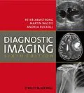
Organised by body system and covering all anatomical regions, Armstrong, Wastie and Rockall:
explain how to interpret images
provide guidelines for interpreting images
discuss common diseases and the signs that can be seen using each imaging modality
illustrate clinical problems with normal and abnormal images
assist diagnosis by covering normal images as well as those for specific disorders
show all imaging modalities used in a clinical context
The authors cover use of plain film, ultrasound, computed tomography, magnetic resonance imaging, radionuclide imaging and interventional radiology, with high quality illustrations and images.
What's new for the 6th edition?
Additional new sections and expanded sections, following reviewer feedback
Updated throughout to ensure recommendations and illustrations reflect modern ultrasound CT, MRI, and nuclear medicine (including PET) practice
Key points and bullet points to aid learning
Autorentext
Professor Peter Armstrong is Professor of Radiology at the Medical College of the St Bartholomew's and the Royal London Hospitals.
Professor Martin Wastie is Professor of Radiology at the University Hospital in Kuala Lumpur, Malaysia. He is formerly a Consultant Radiologist at University Hospital, Nottingham.Dr Andrea Rockall is a Senior Lecturer and Consultant Radiologist at St. Bartholomew's Hospital, London.
Inhalt
Preface.
Acknowledgements.
1 Technical Considerations.
2 Chest.
3 Plain Abdomen.
4 Gastrointestinal Tract.
5 Hepatobiliary System, Spleen and Pancreas with the assistance of Dr Andrew Hatrick.
6 Urinary Tract.
7 Female Genital Tract.
8 Peritoneal Cavity and Retroperitoneum.
9 Bones with the assistance of Dr Kasthoori Jayarani.
10 Joints with the assistance of Dr Kasthoori Jayarani.
11 Spine.
12 Skeletal Trauma.
13 Skull and Brain.
14 Sinuses, Orbits and Neck.
15 Vascular and Interventional Radiology with the assistance of Dr Andrew Hatrick.
Appendix: CT Anatomy of Abdomen.
Index.
