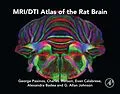
MRI/DTI Atlas of the Rat Brain addresses the MRI/DTI resolution/contrast obtained at the Duke Center for In Vivo Microscopy, which has surpassed that of any other lab by nearly 400x, with images that are satisfactory for the identification of 80%+ of structures previously labeled in Rat Brain in Stereotaxic Coordinates.
This new atlas, from the best imaging/cartography team working in neuroscience today, fully complements the work in Rat Brain in Stereotaxic Coordinates, and will become a new landmark contribution to neuroanatomy as the first and only truly reliable MRI/DTI atlas of the rat brain.
- Ninety-six coronal levels from the olfactory bulb to the pyramidal decussation are depicted
- Delineations primarily made on the basis of direct observations on the MRI contrasts
- Each of the 96 open book pages displays four items- top left the directionally colored fractional anisotropy image derived from DTI (DTI - FAC), top right the diffusion-weighted image (DWI), bottom left the gradient recalled echo (GRE), and bottom right the diagram. The diagram is the synthesis of the information derived from these three images and the two additional images, which we do not display (ARDC and RD). This is repeated for 96 coronal levels, which makes the levels 250 ?m apart.
- The FAC images are shown in full color
- The orientation of sections corresponds to that in Paxinos and Watson's The Rat Brain in Stereotaxic Coordinates (2014)
- The images have been obtained from 3D isotropic population averages (number of rats=5). All abbreviations of structure names are identical to the Paxinos & Watson histologic atlas
Professor George Paxinos, AO (BA, MA, PhD, DSc) completed his BA at The University of California at Berkeley, his PhD at McGill University, and spent a postdoctoral year at Yale University. He is the author of almost 50 books on the structure of the brain of humans and experimental animals, including The Rat Brain in Stereotaxic Coordinates, now in its 7th Edition, which is ranked by Thomson ISI as one of the 50 most cited items in the Web of Science. Dr. Paxinos paved the way for future neuroscience research by being the first to produce a three-dimensional (stereotaxic) framework for placement of electrodes and injections in the brain of experimental animals, which is now used as an international standard. He was a member of the first International Consortium for Brain Mapping, a UCLA based consortium that received the top ranking and was funded by the NIMH led Human Brain Project. Dr. Paxinos has been honored with more than nine distinguished awards throughout his years of research, including: The Warner Brown Memorial Prize (University of California at Berkeley, 1968), The Walter Burfitt Prize (1992), The Award for Excellence in Publishing in Medical Science (Assoc Amer Publishers, 1999), The Ramaciotti Medal for Excellence in Biomedical Research (2001), The Alexander von Humbolt Foundation Prize (Germany 2004), and more.
MRI/DTI Atlas of the Rat Brain offers two major enhancements when compared with earlier attempts to make MRI/DTI rat brain atlases. First, the spatial resolution at 25µm is considerably higher than previous data published. Secondly, the comprehensive set of MRI/DTI contrasts provided has enabled the authors to identify more than 80% of structures identified in The Rat Brain in Stereotaxic Coordinates. - Ninety-six coronal levels from the olfactory bulb to the pyramidal decussation are depicted - Delineations primarily made on the basis of direct observations on the MRI contrasts - Each of the 96 open book pages displays four items- top left, the directionally colored fractional anisotropy image derived from DTI (DTI - FAC); top right, the diffusion-weighted image (DWI); bottom left, the gradient recalled echo (GRE); and bottom right, a diagrammatic synthesis of the information derived from these three images plus two additional images, which are not displayed (ARDC and RD). This is repeated for 96 coronal levels, which makes the levels 250 m apart - The FAC images are shown in full color - The orientation of sections corresponds to that in Paxinos and Watson's The Rat Brain in Stereotaxic Coordinates, 7th Edition (2014) - The images have been obtained from 3D isotropic population averages (number of rats=5). All abbreviations of structure names are identical to the Paxinos & Watson histologic atlas
Autorentext
Professor George Paxinos, AO (BA, MA, PhD, DSc) completed his BA at The University of California at Berkeley, his PhD at McGill University, and spent a postdoctoral year at Yale University. He is the author of almost 50 books on the structure of the brain of humans and experimental animals, including The Rat Brain in Stereotaxic Coordinates, now in its 7th Edition, which is ranked by Thomson ISI as one of the 50 most cited items in the Web of Science. Dr. Paxinos paved the way for future neuroscience research by being the first to produce a three-dimensional (stereotaxic) framework for placement of electrodes and injections in the brain of experimental animals, which is now used as an international standard. He was a member of the first International Consortium for Brain Mapping, a UCLA based consortium that received the top ranking and was funded by the NIMH led Human Brain Project. Dr. Paxinos has been honored with more than nine distinguished awards throughout his years of research, including: The Warner Brown Memorial Prize (University of California at Berkeley, 1968), The Walter Burfitt Prize (1992), The Award for Excellence in Publishing in Medical Science (Assoc Amer Publishers, 1999), The Ramaciotti Medal for Excellence in Biomedical Research (2001), The Alexander von Humbolt Foundation Prize (Germany 2004), and more.
Leseprobe
Key Features
- Ninety-six coronal levels from the olfactory bulb to the pyramidal decussation are depicted. - Delineations primarily made on the basis of direct observations on the MRI contrasts. - Each of the 96 open book pages displays four items- top left the directionally colored fractional anisotropy image derived from DTI (DTI - FAC), top right the diffusion-weighted image (DWI), bottom left the gradient recalled echo (GRE), and bottom right the diagram. The diagram is the synthesis of the information derived from these three images and the two additional images, which we do not display (ARDC and RD). This is repeated for 96 coronal levels, which makes the levels 250 mim apart. - The FAC images are shown in full color. - The orientation of sections corresponds to that in Paxinos and Watson's The Rat Brain in Stereotaxic Coordinates (2014). - The images have been obtained from 3D isotropic population averages (number of rats = 5). All abbreviations of structure names are identical to the Paxinos & Watson histologic atlas. Reproduction of Atlas Figures in Other Publications
As authors, we give permission for the reproduction of any figure from the atlas in other publications, provided that the atlas is cited. Permission from the publisher may be sought directly on-line via the Elsevier homepage (http://elsevier.com/locate/permissions) or from Elsevier Global Rights Department in Oxford, UK: phone: (+44) 1865843830, fax: (+44) 1865 853333, email: permissions@elsevier.com. Acknowledgments
This work was supported by NHMRC grant 1086643, the Australian Research Council Centre of Excellence for Integrative Brain Function (ARC Centre Grant CE140100007). All MR images were acquired at the Duke Center for In Vivo Microscopy, an NIH/NIBIB Biomedical Technology Resource Center (P41 EB015897). AB gratefully acknowledges support from NIH through K01AG041211. We thank Dr. Emma Schofield and Mark Foster for expert assistance in the organization of image sets. Introduction
The development…